
👁️ What Is Actually Inside Your Eye?
Yesterday, you learned something important:
Seeing is not as simple as opening your eyes.
Light has to enter the eye, travel through several structures, reach the retina, and eventually create signals that travel toward the brain.
But that creates another question:
What exactly is inside an eyeball?
Soon, you are going to find out firsthand.
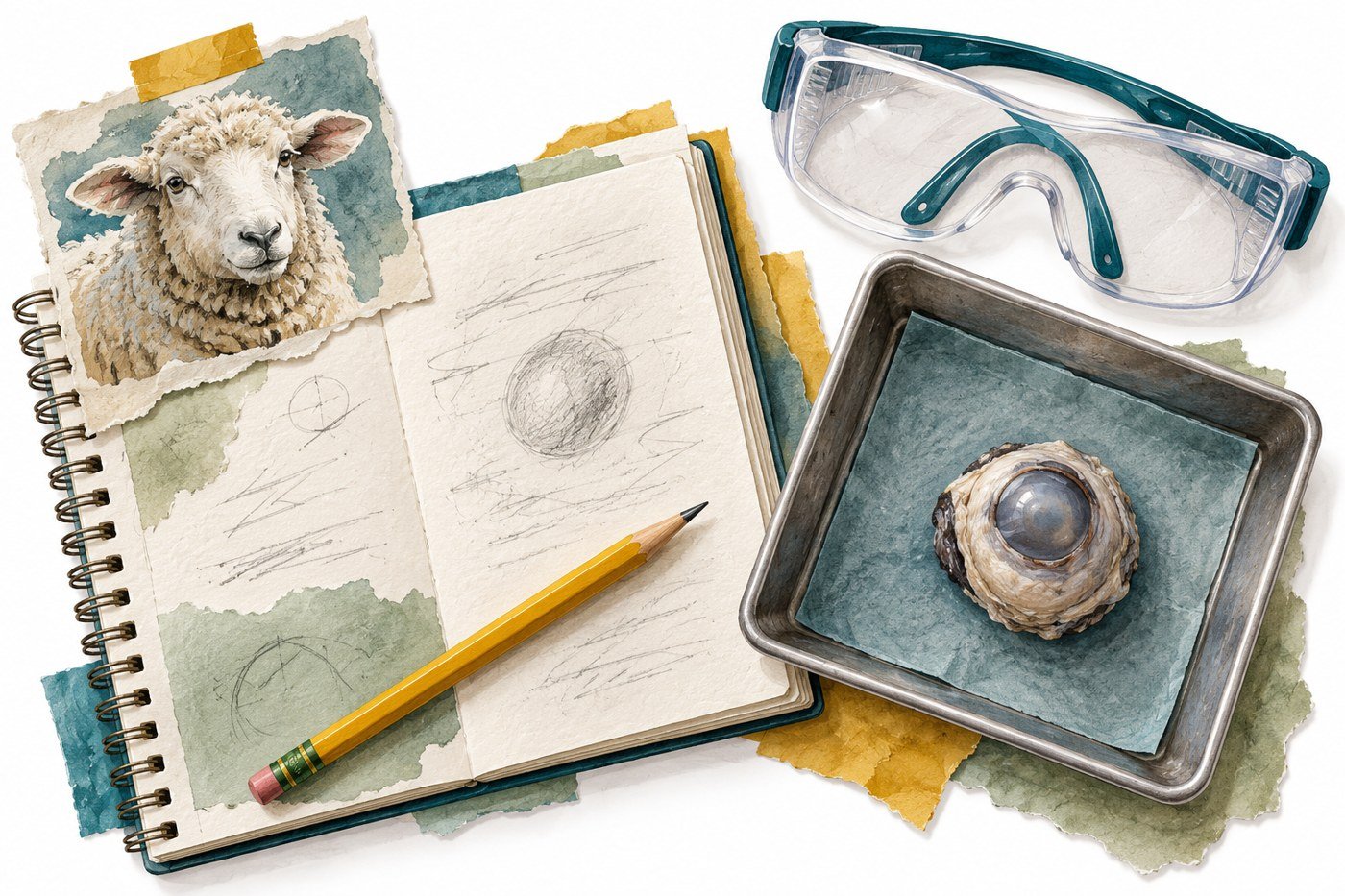
You will be dissecting a sheep eye.
Before anyone starts cutting into an actual eyeball, however, it would probably be useful to know what you are hoping to find.
So today, we are taking a trip through the eye.
🛡️ 1. Sclera: The Protective Outside
Touch the white part of your eye.
Not literally right now. Keep your fingers out of your eyes.
That white outer layer is called the sclera.
The sclera is a tough outer coating that helps protect the eye and maintain its shape.
When we examine a sheep eye, the sclera will be one of the easiest structures to recognize.
🔍 2. Cornea: The Clear Window
At the front of the eye is the cornea.
Unlike the white sclera, the cornea is normally transparent.
Why?
Because light needs to pass through it.
Its curved surface also bends, or refracts, incoming light and begins the focusing process.
Think of the cornea as the eye’s front window.
🎨 3. Iris: The Colored Part
Brown eyes?
Blue?
Green?
Hazel?
The colored structure you see is the iris.
But the iris does much more than give people different eye colors.
It controls the size of the pupil and therefore helps regulate how much light enters the eye.
⚫ 4. Pupil: The Opening
The black circle in the middle of your iris is the pupil.
Remember from Post 1:
The pupil is an opening, not a solid black structure.
Light passes through this opening on its way farther into the eye.
🔎 5. Lens: Fine-Tuning the Focus
Behind the iris is the lens.
The lens is clear and helps focus incoming light onto the retina.
The lens can change shape, allowing the eye to focus on objects at different distances.
You will actually be able to remove the lens when you dissect the sheep eye.
And unlike many microscopic structures we discuss in science, this one is large enough to hold.
Science finally gives you something cooperative.
💧 6. Aqueous and Vitreous Humor
Your eyeball is not empty.
Much of its interior contains clear fluids or gel-like material.
The aqueous humor is a watery fluid found toward the front of the eye.
The vitreous humor is a clear, gel-like substance filling much of the large space behind the lens. The National Eye Institute describes vitreous humor as the clear gel filling the inside of the eye.
This material helps the eyeball maintain its shape while still allowing light to pass through.
📡 7. Retina: The Light Detector
At the back of the eye is the retina.
This is one of the most important structures in vision.
The retina is light-sensitive tissue containing cells called photoreceptors.
Photoreceptors respond to light and begin turning visual information into electrical signals.
Without a functioning retina, simply focusing light correctly would not be enough to produce normal vision.
🧠 8. Optic Nerve: The Connection to the Brain
Finally, visual signals need somewhere to go.
The optic nerve carries information from the retina toward the brain.
The human optic nerve contains more than one million nerve fibers.
Think of it as a massive information cable connecting your eye to your brain.
🛣️ Follow the Light
Try to follow the path light takes:
Cornea → Pupil → Lens → Vitreous humor → Retina
Then visual information leaves the retina through a different pathway:
Retina → Optic nerve → Brain
Those are two related but different paths.
One describes the movement of light.
The other describes the movement of visual information.
That difference is important.
🔬 Vocabulary Check
You should now recognize:
Sclera – tough outer covering
Cornea – clear front surface that helps bend light
Iris – colored structure that controls pupil size
Pupil – opening through which light enters
Lens – clear structure that focuses light
Vitreous Humor – clear gel inside the eye
Retina – light-sensitive tissue at the back of the eye
Optic Nerve – carries visual signals toward the brain
✍️ Your Blog Response
Imagine you have been shrunk to the size of a dust particle and are traveling through an eyeball along with a beam of light.
Write 6–8 complete sentences describing your journey.
You must correctly describe and use:
cornea • pupil • lens • retina • optic nerve
Your response must also explain:
Why wouldn’t focusing light perfectly onto the back of the eye be enough for you to see if the retina were not working?
Use evidence from today’s reading to support your explanation.
When you’re finished, check out the rest of our blog for more tips, ideas, and activities to help you learn and grow. Be sure to follow our classroom Instagram page for behind-the-scenes moments, project highlights, and fun updates. Let’s work together to make learning fun, exciting, and something you look forward to every day!


34 Responses
A cornea is a clear front surface that helps bend light.A pupil is a opening through which light enters.A lens is a clear structure that focuses light.A retina is light sensitive tissue at the back of the eye.An optic nerve carries visual signals toward the brain.They all work together by going in steps for the eye to work.
If I shrunk to a dust size particle and entered an eye, I would see light bending from the clear layer called the cornea. Then I would see the light passing through the pupil and into the lens helping by focusing the light. After that I would see the light pass through the clear gel like liquid called the vitreous humor. And it would go into the retina and send signals through the optic nerves. allowing us to see.
Focusing light will sometimes not work because sometimes the cooler of your eye might be the problem because the light has to go though the the lens then cornea then the purple then the iris then the retina the the retina connects to the optic nerve that then connects it to the brain and that is why light doesn’t perfectly onto the back of the eye.
The cornea, which is the clear part at the front of your eye. Then the pupil, where the light enters the eye. After that, the lens focused the light so it could make a clear picture. Then light then reaches the retina, which is in the back of the eye and senses the light. The retina turns the light into signals and sends them through the optic nerve to the brain. If the retina was not working, it would not matter if the light was focused perfectly because the retina could not sense it. Without the retina sending signals to the brain, you would not be able to see the image.
Light onto the back of the eye is not enough because the retina is the is responsible for translating that light into electrical signals that the brain can understand.If the retina is not working, the light cannot be converted or processed, resulting in blindness.
The first thing I see is the iris the color is green and a little bit of brown is in there too. I also see the cornea it’s clear, and the surface is curved because it needs light to pass through it, Its curved surface also bends, or refracts incoming light and begins the focusing process. I also noticed the pupil its not a black structure it is an opening, light passes through it to go farther into the eye. And I see the retina, the retina is a light sensitive tissue at the back of your eye. The Vitreous Humor is clear gel in your eye. The Sclera is the tough outer covering. The Lens is a clear structure that focuses light. The Optic nerve carries visual signals towards the brain.
If I shrunk into the size of a dust particle traveling along the eyeball I would first the the pupil. Its an opening so its what I came through, I would then feel light beaming on me through the cornea. After I nearly get burned to a crisp due to the hot heat, I would travel along the iris. Gazing at the beautiful eye color Retina. Which is a sensitive tissue at the back of the eye. I would then feel signals traveling. They’re called Optic Nerves and they help send signals to the brain.
If I shrunk and went inside an eye, I see the eye color is blue. I also see the cornea. Then the pupil, which would be an opening , and light passes though. Then the retina, which is the tissue behind my eye. Then the optic nerve sends signals to my brain, and I can see.
The inside of the eye has several important parts that help us see.The pupil lets light enter, while the lens focuses the light.At the back of the eye, the retina senses the light and sends information to the brain through the optic nerve. The Inside of the eye is also filled with a clear , jelly like substance called vireouse humor
Your eyeball contains several key layers and structures that work together to bend, focus, and convert light into images your brain can see Retina: The light-sensitive layer at the back of the eyeball that changes light into electrical signals.Vitreous Gel: A clear, jelly-like substance that fills the large space behind the lens and keeps the round shape of the eye.Optic Nerve: A thick cable at the back of the eye that sends visual messages from the retina to t The strong white outer wall of the eyeball that protects the inner parts
The first thing I see is the iris the color is yellow and a little bit of orange is in there too. I also see the cornea it’s clear, and the surface is curved because it needs light to pass through it, Its curved surface also bends, or refracts incoming light and begins the focusing process. I also noticed the pupil its not a black structure it is an opening, light passes through it to go farther into the eye. And I see the retina, the retina is a light sensitive tissue at the back of your eye. The Vitreous Humor is clear gel in your eye. The Sclera is the tough outer covering. The Lens is a clear structure that focuses light. The Optic nerve carries visual signals towards the brain
A cornea is the clear part of the front of your eye that helps bend light. The pupil helps open through so that light enters. Then the lens help clear structure that focuses on light. The retina is the sensitive tissue on the back of the corner of the eye. If the tissue breaks then it will probably cause blindness. I think the optic nerve is something that carries visual signals to the brain making the eyes see clearly.
The thing I see is the iris the color is green and a little bit of brown is in it too. I also see the cornea it’s clear, and the surface is curved because it needs light to pass through it, Its curved surface also bends, or refracts incoming light and begins the focusing process. I also noticed the pupil its not a black structure it is an opening, light passes through it to go farther into the eye. And I see the retina, the retina is a light sensitive tissue at the back of your eye. The Vitreous Humor is a clear gel in your eye. The sclera is the tough outer covering. The Lens is a clear structure that focuses light. The Optic nerve carries visual signals towards the brain.
If I shrunk to the size of a dust particle and went inside of an eye then I would first see the cornea which is the front surface and it helps bend light. Then I would be at the pupil of the eye which is the opening where light enters. Then after that I would see the lens which is the clear structure that focuses light and I would also see the Vitreous humor which is the clear gel in the eye.Then at the back of the eye is the retina which is the light sensitive tissue. The Optic Nerve carries the visual signals towards the brain and so that is what makes you able to see.
If I shrunk to a size small enough to enter an eye the first thing that will most likely see is my Iris, because it is the thing that shows me the color and size of my Iris.The next thing I will most likely see is the corona because the clear front surface that blends in with the light.If I keep going to the back of the eye I will see the lens witch is also the clear structure that focuses light. Next I will most likely see the retina the light-sensitive tissue at the back of the eye. The next thing I will see is the optic nerve which is the thing that carries visual signals toward the brain.If I keep going back toward the eye then I will most likely see the brain which connect to eye so that the brain knows what the eyes see.
Focusing light will sometimes well not work because sometimes the cooler of your eye might be the problem because the light has to go though the the lens then cornea then the purple then the iris then the retina the the retina connects to the optic nerve that then connects it to the brain and that is why light doesn’t perfectly onto the back of the eye.
If I shrunk and became a dust particle in an eye first, I would notice a thin white layer with some light reflecting/ bending off of the white layer that is called the cornea. Next through my adventure I would notice the pupil in the middle of the eye the pupil has an opening that lets light enter. After that I would see the lens behind the iris the lens looks like a clear smooth structure the lens work by focusing the light. Then I saw the Vitreous Humor it is a clear gel inside the eye. Afterward I saw the retina the retina is a “light- sensitive tissue at the back of the eye”. Later on I saw the optic nerve which visuals signals to or toward the brain.
First what you would go through is the cornea, a clear front surface that helps bend light and your iris that is the color part on the eye. Next, you would go through the pupil, the pupil is not just a black eye it is actually an opening that light goes through. Next is the Retina . the Retina is light sensitive tissue that is in the back of the eye. You will then go through the lens, the lens is a clear structure that helps focus light. After that the last thing you go through is the optic nerve, the optic nerve carries the visual signals towards the brain. A person would not be able to see if the retina was working fully or not looking at it because it has cells inside that respond to light and starts to turn visual information into electrical signals.
If I were to shrink and see inside an eye, I would observe that it is blue. I can see the cornea as well. Light then enters through the pupil, which would be an aperture. The tissue behind my eye is called the retina. My brain receives information from the optic nerve, allowing me to see.
As a tiny particle of dust traveling with a beam of light, your journey begins when you pass right through the clear front window known as the cornea.
Next, you zoom straight through the dark opening of the pupil and hit the clear lens that fine-tunes your focus. You drift across the clear gel of the vitreous humor before crashing onto the back wall at the retina. Here, light-sensitive photoreceptor cells catch you and convert your energy into a brand-new electrical message. That newly created signal then races away from the eyeball through the optic nerve straight toward the waiting brain.
Why a Working Retina Matters
If I struck into a small size to where I would be able to go inside of the human eye I would first see the iris , the iris is helps you see everything like color , size and etc. if I keep going the next thing that will be seen is the corona . The corona is a clear surface like thing that helps blend lighting . The pupil is not just black it’s actually an opening .
If I were to shrink and first become a speck of dust inside an eye, I would observe a thin white layer—the cornea—where light is reflected or refracted. Continuing my adventure, I would see the pupil at the center of the eye; it features an opening that allows light to enter. Next, I would see the lens behind the iris; it appears as a smooth, transparent structure, and its function is to focus light. Then I would see the vitreous humor, a transparent gel located inside the eye. Following that, I would observe the retina, a light-sensitive tissue located at the back of the eye. Finally, I would see the optic nerve, which transmits visual signals to the brain.
If I shrunk into the size of a dust particle I will see the cornea first which helps the eye to bend light. Then the Pupil is where the opening where lights pass through. After that I would see the Lens which allows the eye to focus on objects from different distances and also is a clear structure that focuses light. Then I see the Vitreous Humor which has a clear gel inside the eye. Then I also see the Retina which is a light sensitive tissue at the back of the eye.Lastly I passes through the Retina then see the Optic Nerve which carries visual signals toward the brain.
The cornea is the clear front surface of your eye. Then the pupil, the hole through which light enters into the eye. Then the lens brought the light together to form a clear picture. Then light reaches the retina, the light-sensitive layer at the back of the eye. The retina converts the light into signals and sends them through the optic nerve to the brain. If the retina was not functioning, then it would not matter if the light was focused perfectly because the retina would not be able to sense it. If the retina is not sending signals to the brain, you would not be able to see the image.
If I shrunk into the size of a dust particle traveling along the eyeball I would first the the pupil. Its an opening so its what I came through, I would then feel light beaming on me through the cornea. After I nearly get burned to a crisp due to the hot heat, I would travel along the iris. Gazing at the beautiful eye color Retina. Which is a sensitive tissue at the back of the eye. I would then feel signals traveling. They’re called Optic Nerves and they help send signals to the brain.
The cornea is a clear surface of your eyes. Then the pupil, is a hole which light enters the eyes. Then the lens bring the light together to make your vision clear. The light goes to the retina a layer behind the back of our eyes, The retina gives signals and sends them through an optic nerve to the brain. So that is how our eyes work.
Light has to enter the eye, travel through several structures, reach the retina, and eventually create signals that travel toward the brain white part of you eye is That white outer layer is called the sclera is a tough outer coating that helps protect the eye and maintain its shape At the front of the eye is the cornea Because light needs to pass through it.
My journey begins as I ride a beam of light through the clear, protective cornea, which shrinks the light to start focusing our path. Next, we go straight through the pupil, the dark hole starts opening that goes to the right size to let us pass into the inner eye. right after, we crash through the lens, which changes its shape to focus our beam. We then fly across the fluid-filled of the eye and land on the retina, the sensitive part starts lining the very back of the eyeball. Here, Finally, we travel along the nerve to the brain, where the final image is actually seen and understood.
Light has to enter the eye, travel through several structures, reach the retina, and eventually create signals that travel toward the brain. White part of you eye is That white outer layer is called the sclera is a tough outer coating that helps protect the eye and maintain its shape .At the front of the eye is the cornea Because light needs to pass through it.The colored structure you see is the iris but the iris does much more than give people different eye colors it controls the size of the pupil and therefore helps regulate how much light enters the eye. The black circle in the middle of your iris is the pupil. Behind the iris is the lens the lens is clear and helps focus incoming light onto the retina The lens can change shape allowing the eye to focus on objects at different distances. Much of its interior contains clear fluids or gel-like material the aqueous humor is a watery fluid found toward the front of the eye the vitreous humor is a clear, gel-like substance filling much of the large space behind the lens The National Eye Institute describes vitreous humor as the clear gel filling the inside of the eye This material helps the eyeball maintain its shape while still allowing light to pass through.
The human eye is a complex, spherical organ protected on the outside by the sclera—the tough, white layer that gives it its shape. At the front lies the cornea, a curved, transparent structure that acts as the eye’s first lens, allowing light to pass through and protecting the iris. Directly behind the cornea is the anterior chamber, filled with a clear fluid called aqueous humor, which nourishes the surrounding tissues and maintains the internal pressure of the eyeball.
Focusing light perfectly onto the back of the eye is not enough to see because the retina is responsible for converting light into electrical signals that the brain can understand. If the retina is not working, the light cannot be processed or transmitted to the brain.
First coming to a beam of light, the journey begins as we go to the cornea, the clear protective outer window that bends a inward path. Next, we go through the pupil, the dark adjustable opening that controls how much light enters the inner chamber of the eye. Immediately behind it, we pass through the crystal-clear lens, which actively reshapes itself to focus our beam perfectly toward the back of the eye. We now go into the retina, the complex layer lining the rear wall where millions of light-sensitive receptor cells await. However, simply focusing light perfectly onto the back of the eye is not enough to actually see if the retina is damaged or non-functional. The eye requires the retina to act as a vital converter, turning that focused light energy into electrical nerve impulses. Without this conversion, no messages can travel along the optic nerve to the brain, which is the organ that ultimately interprets these signals as a visual image.
Getting shrunk and then going though the eye. You would first see the cornea the cornea is normally transparent. because light needs to pass through it. Its curved surface also bends, or refracts, incoming light and begins the focusing process. Think of the cornea as the eye’s front window. then you would find the pupil the pupil is a oping not a solid object a pupil. near the pupil is the iris that can be different colors all having there own ability. the you would be up against the lens. The lens is clear and helps focus incoming light onto the retina.The lens can change shape, allowing the eye to focus on objects at different distances.You will actually be able to remove the lens when you dissect the sheep eye.And unlike many microscopic structures.then next in line is the retina when light comes though. it also connects to the. optic nerve carries information from the retina toward the brain.The human optic nerve contains more than one million nerve fibers.Think of it as a massive information cable connecting your eye to your brain. and that is the end of the trip.
Our eyes contain a complex network of tissues,fluids and specialized cells that work together to capture light.
. cornea: A clear,dome-shaped window at very front that bends light as it enters.
.irus:The colorful muscle ring that controls;s how much light gets in
. pupil:The dark opening in the middle of the irus that gets bigger in the dark and smaller in bright light
.sclera: The thought white outer coating that protects the eyeball